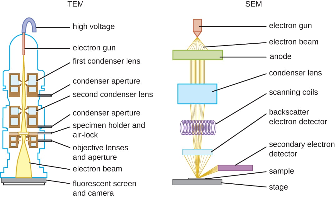
Source of Radiation: A key difference lies in the type of radiation used; light microscopes use light, while electron microscopes use electrons.
Wavelength and Resolution: Electron microscopes have a much shorter wavelength than light, resulting in a higher resolution. The resolution of electron microscopes is 0.5nm, compared to 200nm for light.
Magnification: While both types can magnify specimens, electron microscopes offer higher magnification due to their superior resolution.
Lenses: Light microscopes use glass lenses to focus light, whereas electron microscopes use electromagnets to focus the electron beam.
Specimen condition: Light microscopes can be used to view living specimens whereas electron microscopes can't as the specimen must be in a vacuum.
SEM vs TEM
There are two types of electron microscopes, TEMs and SEMs.
Transmission Electron Microscope (TEM): The electron beam passes through the specimen, allowing visualization of thin sections and internal cell structures.
Scanning Electron Microscope (SEM): The electron beam scans the surface of the specimen, providing a 3D appearance and allowing observation of surface structures. However, SEMs have a lower resolution compared to TEMs, ranging from 3 nm to 20 nm.
How do we know which type of microscope to use?
=
We use a light microscope when we need living cells, colour is required or measurements using the eyepiece graticule are needed.
We use TEMs when we need to study very small structures within a cell, such as ribosomes, endoplasmic reticulum, and membranes.
We use SEMs when we need to view the surface of a structure in 3D.
We use both SEMs and TEMs to study viruses as they are too small to be viewed with a light microscope.